Pupa Gilbert earns WARF Named Professorship

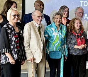
WARF named professorship award ceremony
Front row from left: Dorota Brzezińska (UW-Vice Chancellor for Research), John D. Wiley (former UW Chancellor), Pupa Gilbert (John D. Wiley Professor of Physics), Jennifer Mnookin (UW Chancellor)
Back row from left: Lauren Bishop (professor of Social Work, Romnes winner), Charles Lee Isbell, Jr. (UW Provost), Susan Harness (Maria Stuchly Professor of Electrical Engineering), Russ Shafer-Landau (Elliott Sober Professor of Philosophy).

Physics professor Pupa Gilbert is one of eight UW–Madison faculty awarded WARF Named Professorships, which come with $100,000 and honor faculty who have made major contributions to the advancement of knowledge, primarily through their research endeavors, but also as a result of their teaching and service activities. Award recipients choose the names associated with their professorships.

Gilbert, the John D. Wiley professor of physics, joined the Department of Physics in 1999 as a full professor, following staff scientist positions in the Italian National Research Council (CNR) in Rome and the Swiss Institute of Technology in Lausanne (EPFL). Her research focuses on biomineralization, that is, on understanding the nanoscale formation mechanisms of natural biominerals, especially coral skeletons, but on a side she also studies sea urchin spines, mollusk shell nacre, tooth enamel, and inner ear cochleaer bone. She also strives to understand the evolution of biomineralization, and how biominerals interact with the environment in the past, present, and future. She is actively working to save coral reefs from climate change.

Her role in the profession has included being research director for the NSF-funded Synchrotron Radiation Center at UW–Madison, chair of the Division of Biological Physics (DBIO) of the American Physical Society, a Radcliffe Fellow at Harvard University, and establishing “the Cnidarians,” a group of tens of diverse undergraduate students at UW–Madison who produce great discoveries by doing massively parallel data processing. She teaches Physics in the Arts, a popular course for non-science majors on light, color, and sound. She was knighted by Italian President Carlo A. Ciampi in 2001.

“Prof. Pupa Gilbert is a luminary in the field of biomineralization, developing a fundamental understanding of these crucial processes that are shared by many diverse organisms,” says Kevin Black, chair of the UW–Madison department of physics. “Prof. Gilbert is a knight of the Italian Republic, and stalwart advocate for involving undergraduates, in particular those from underrepresented groups in her research. This award recognizes the many contributions to the academy in her long and productive interdisciplinary research program and is well deserved.”

For her named professorship, Gilbert chose John D. Wiley, the former UW–Madison Chancellor who earned his MS and PhD from this department. He was a member of the technical staff at Bell Laboratories, then a visiting scientist at Max Planck Institute for Semiconductor Physics. He joined the faculty at UW–Madison as professor of electrical and computer engineering in 1975, where he did groundbreaking research on semiconductors and metals, and earned major patents at WARF. Later, he became Chair of the Materials Science Program, Dean of the Graduate School, Provost, and then Chancellor (2001-2008), all at UW–Madison.

Wiley also started campus-wide initiatives such as cluster hiring and originated many campus building projects. He was the first Director of the Wisconsin Institute of Discovery, a position he held from 2008 until he retired in 2011.

Gilbert chose Wiley, whom she calls “a brilliant physicist,” for her named professorship for all the reasons listed above. She also credits him with bringing her to UW–Madison.

“When there was a cluster hire in biophotonics back in 1999, John Wiley wanted me to join the university and he had them include the word spectromicroscopy — the synchrotron method that I developed — into the search to encourage me to apply,” Gilbert says. “I really didn’t want to apply, I was on my way to CalTech, but of course I couldn’t say no to John.”

In total, thirty-two University of Wisconsin–Madison faculty were awarded fellowships from the Office of the Vice Chancellor for Research for 2024-25, including eight WARF named professorships, 13 Romnes faculty fellowships, and 11 Kellett mid-career awards. The awardees span the four research divisions on campus: arts and humanities, physical sciences, social sciences and biological sciences.

“These awards recognize excellence in faculty research, academics, and outreach at various stages of their scholarly careers and provide an opportunity for continued development of their research programs,” says Cynthia Czajkowski, interim vice chancellor for research. “I look forward to seeing the results of their imaginative use of these funds.”

The awards are possible due to the research efforts of UW–Madison faculty and staff. Technology that arises from these efforts is licensed by the Wisconsin Alumni Research Foundation and the income from successful licenses is returned to the OVCR, where it’s used to fund research activities and awards throughout the divisions on campus.
Pupa Gilbert earns undergraduate mentoring award

Congrats to Prof. Pupa Gilbert on earning an Award for Mentoring Undergraduates in Research, Scholarly and Creative Activities!

The Office of the Provost offers awards to recognize the important role mentors play in fostering undergraduates’ intellectual, personal and professional growth through participation in high-impact practices including research, scholarly and creative endeavors. These awards provide mentors with recognition for their excellence in mentoring undergraduates and their contribution to our students’ Wisconsin Experience.

Gilbert has mentored many undergraduate and graduate students as a physics professor at UW–Madison. Some of these students, like Andrii Hopanchuk ‘24, worked on individual research projects and served as an undergraduate teaching assistant for her popular course, Physics 109, Physics in the Arts.

“Pupa has supported me in exploring my own work, which ultimately culminated in me single-authoring a paper and publishing it in a peer-reviewed journal,” Hopanchuk wrote in his nomination letter. “Another way Pupa has impacted my professional growth was by maintaining a high standard for me (I wish more people did that).”

In 2020, when in-person undergraduate student research positions were almost non-existent, Gilbert had an idea to hire over a dozen students, largely from the Mercile J. Lee Scholars program, to conduct remote analysis on complicated data sets. Machine learning models are only as good as the data they are trained on, and Gilbert reasoned that a large group of diverse students conducting parallel analyses would yield conclusions that computers never could.

This group of 15 students, dubbed the Cnidarians (from the phylum that includes corals, anemones, and jellyfish — animals these students analyzed in Gilbert’s group), each processed the data, making 11 distinct decisions along the way. At weekly Zoom meetings, results were compared. If the majority of Cnidarians agreed on a solution, it was likely the correct one; if not, the students walked others through the process of how they made the decisions they did.

“We (the Cnidarians, really!) discovered many ways to improve data processing,” Gilbert wrote in a Springer Nature “Behind the Paper” feature. “Diversity is our strength. The accuracy, precision, and overall quality of the results is outstanding and unprecedented.”

Many Cnidarians were among Gilbert’s nominators for the Mentoring award.

“Pupa is one of the few professors I have experienced whose energy for learning is contagious – any student under her mentorship is bound for success,” wrote Virginia Quach ’23, now a Pharmacy doctoral student.

Isabelle LeCloux is a senior chemical engineering major who worked with Gilbert both at UW–Madison and at Lawrence Berkeley National Lab this past summer. She wrote:

“Working with other undergraduates in the welcoming and encouraging environment fostered by Professor Gilbert has allowed me to explore topics that I otherwise would not have been exposed to in my curriculum. Her positive attitude makes her a delight to work with and I always feel that I can ask any questions I may have regarding our work in the group.”

Biology major CP Breit joined the Cnidarians after they had already been working together for a year, but she said that Gilbert made it easy to join the already-established team.

Breit wrote: “During this training period Dr. Gilbert was incredibly supportive and really gaveme the confidence to start processing data quickly. There were certainly shortcomings in my technical acumen but Dr. Gilbert was always positive and encouraging, allowing me to make mistakes, learn how to correct them, and improve.”

All the Cnidarians were named as co-authors on the Gilbert group’s most recent paper, published in February in the prestigious journal Nature Communications. Other Cnidarians co-authored a 2022 JACS paper.

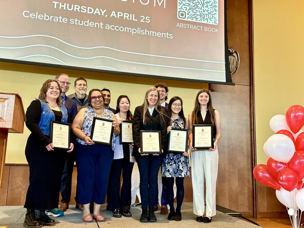
Gilbert and other mentoring awardees were recognized at the Mentor Awards Ceremony held during the annual Undergraduate Research Symposium on April 25.

a group of people stand on a stage, all holding framed certificates
The 2024 Mentor award winners at the Undergraduate Research Symposium. Pupa Gilbert is in the front row, third from right (photo provided by Pupa Gilbert)

Fringe benefits: new technique makes x-rays more laser-like

Detecting a chemical change is often easy: colors may change, heat may be released, or something may smell different. Seeing reactions at the molecular level is not quite so easy, but knowing exactly when and how chemical bonds form or atoms move around is crucial to understanding chemical processes.

animated gif showing atomic-level structural changes taking place during the chemical reaction
X-ray pulses allow researchers to create real-time movies of chemical reactions at the molecular scale (click on image to see animation)

In a new study published March 15 in Proceedings of the National Academy of Sciences, University of Wisconsin–Madison physics professor Uwe Bergmann and his collaborators have turned ultrafast x-ray pulses into something more akin to an optical laser, with cleaner, directional pulses. Their work may lead to visualizing chemical reactions faster than ever at the atomic scale.

“This work is the first step to do with x-rays the same kind of [techniques] which you do with regular lasers,” says Bergmann, the study’s senior author. “We have opened a time window for looking at chemical processes with attosecond [one billionth of one billionth of a second] precision. It’s a new frontier.”

Profile picture of Uwe Bergmann
Uwe Bergmann

Around a decade ago, researchers began using powerful x-ray free-electron lasers, which allow them to make ultrafast movies of molecular changes in real time on the femtosecond scale (one thousand times slower than an attosecond). Compared to visible lasers, which provide clean, single-wavelength beams of light, x-ray lasers are somewhat dirty: they contain multiple wavelengths of light of randomly varying intensity.

“What all scientists have done, and are still doing, is that you just adapt to what you get and then you design your experiments around them,” Bergmann says. “That also means that certain experiments, which in the optical laser regime are now standard, have not been possible.”

Bergmann and his colleagues somewhat accidentally discovered a way to make x-rays more like an optical laser. In their experimental setup, they shine intense but dirty x-ray pulses at a manganese sample. When these pulses hit a manganese atom, a lower-level electron is ejected and the hole it leaves is rapidly filled by a higher-level electron. The energy difference is emitted as a photon of a characteristic color. Very intense pulses can create enough of these holes. An emitted photon can effectively stimulate the emission of another one, leading to an avalanche of stimulated X-ray emission, mostly in the forward direction towards their detector.

Every so often, the detector that captures these stimulated emissions showed something they were not expecting: strong fringes, the characteristic pattern that results from constructively and destructively interfering signals. The fringes suggested that they had observed two x-ray emission pulses, separated by only a few femtoseconds.

“We were confused,” Bergmann says. “How did such a pair of x-ray pulses come about?”

After many discussions, calculations, and simulations, the team ruled out many possible explanations, until they finally realized what had happened: occasionally, two of the many spikes in the dirty pulses were much stronger than the rest of them. When these strong spikes occurred a few femtoseconds apart, and each had enough intensity, a clean pair of stimulated x-ray emission pulses emerged.

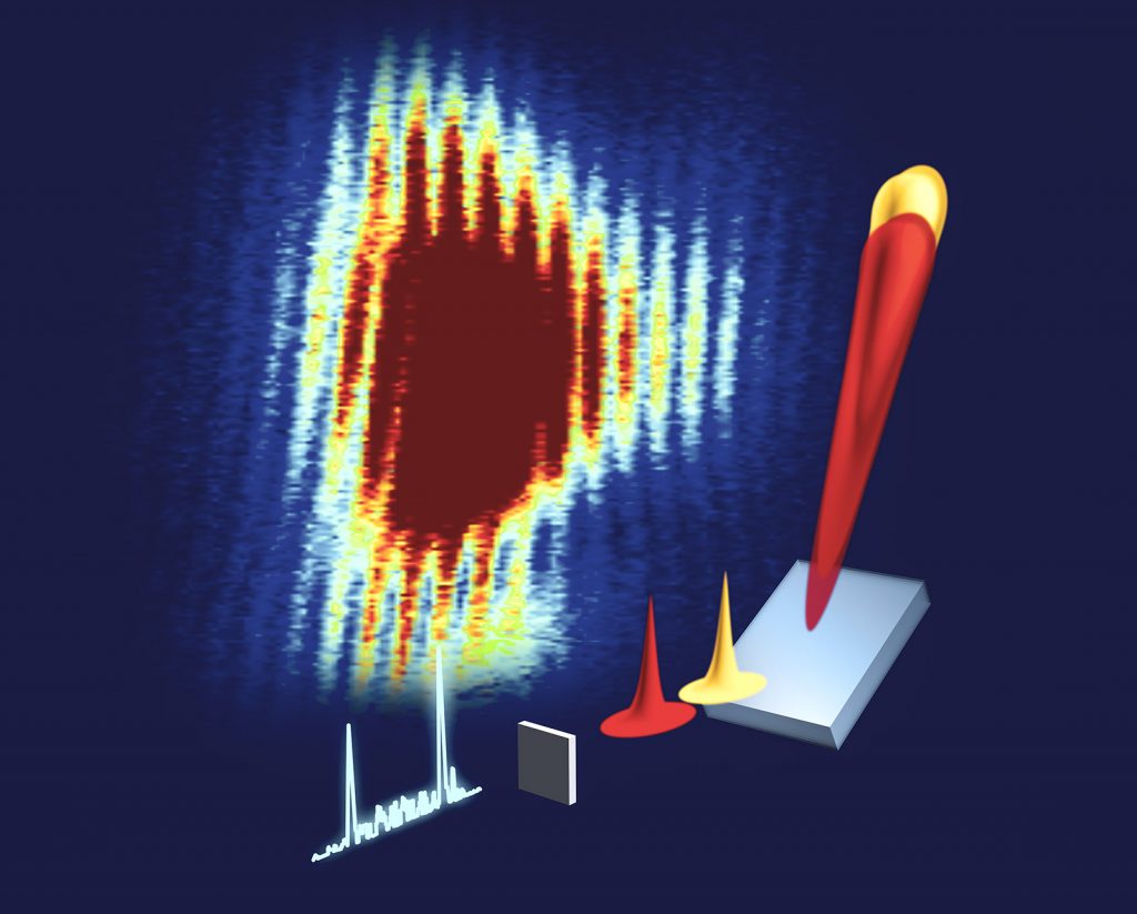
depiction of the experiment: a pulse of x-rays is shown as jagged white peaks. that pulse travels through the sample, represented as a thin square, where the signals are stimulated into larger, unidirectional peaks, shown as a red and yellow taller peaks. those peaks are streched out length-wise by the monochromator such that they now overlap. The result is a rainbow-colored splotch representing intensity of the measured signal. The splotch looks like a comb, which are the fringes.
Two strong x-ray pulses hit the manganese sample, are stretched through the monochromator, and overlap, leading to the characteristic fringes.

Before these two pulses reach the detector, they first travel through a monochromator — essentially a prism that stretches light, much like how white light passing through a clear prism is stretched into a rainbow. These stretched pulses then overlap timewise, and those frequencies that are in phase with each other can add up to become more intense or cancel each other out into dark troughs. Hence, the fringes.

“At times, each signal is rather clean and of similar strength, and one obtains very strong interference fringes,” Bergmann says. “We know that the fringe spacings are directly related to the time difference of the two pulses, and because we can measure them very precisely, we can obtain their time difference with extreme, attosecond precision.”

Currently, these pulse pairs are generated very rarely, Bergmann and his collaborators will work with the accelerator scientists to find ways to manipulate the ‘dirty’ pulses and enhance the chance of producing the pairs. They are optimistic that their work opens the door to new applications of such x-ray pulse pairs — the types of techniques that are used commonly with visible laser light.

“We’re trying to move nonlinear laser optics into the x-ray regime,” Bergmann says. “In the x-ray regime, you can probe certain phenomena that you just cannot optically access. X-ray wavelengths are comparable to the distances between atoms, and we can knock out lower-level electrons to get element specificity with them.”

Bergmann’s contribution to this research was funded in part by the U.S. Department of Energy, Laboratory Directed Research and Development program at SLAC National Accelerator Laboratory (DE-AC02-76SF00515). Other authors were supported by various funding as described in the study.

Coral skeleton formation rate determines resilience to acidifying oceans

A new University of Wisconsin–Madison study has implications for predicting coral reef survival and developing mitigation strategies against having their bony skeletons weakened by ocean acidification.

Though coral reefs make up less than one percent of the ocean floor, these ecosystems are among the most biodiverse on the planet — with over a million species estimated to be associated with reefs.

The coral species that make up these reefs are known to be differently sensitive or resilient to ocean acidification — the result of increasing atmospheric carbon dioxide levels. But scientists are not sure why.

In the study, researchers show that the crystallization rate of coral skeletons differs across species and is correlated with their resilience to acidification.

A woman holding two coral species stands in front of a body of water
“Finding solutions that are science-based is a priority,” says physics professor Pupa Gilbert, shown here with samples of scleractinian coral along the Lake Monona shoreline in Madison. | Photo: Jeff Miller

“Many agencies keep putting out reports in which they say, ‘Yes, coral reefs are threatened,’ with no idea what to do,” says Pupa Gilbert, a physics professor at UW–Madison and senior author of the study that was published Jan. 17 in the Journal of the American Chemical Society. “Finding solutions that are science-based is a priority, and having a quantitative idea of exactly what’s happening with climate change to coral reefs and skeletons is really important.”

Reef-forming corals are marine animals that produce a hard skeleton made up of the mostly insoluble crystalline material aragonite. Aragonite forms when precursors made up of a more soluble form, amorphous calcium carbonate, are deposited onto the growing skeleton and then crystallize.

The team studied three genera of coral and took an in-depth look at the components of their growing skeletons. They used a technique that Gilbert pioneered called PEEM spectromicroscopy, which detects the different forms of calcium carbonate with the greatest sensitivity to date.

When they used these spectromicroscopy images to compare the thickness of amorphous precursors to the crystalline form, they found that Acropora, which is more sensitive to acidification, had a much thicker band of amorphous calcium carbonate than Stylophora, which is less sensitive.

A third genus of unknown sensitivity, Turbinaria, had an even thinner amorphous precursor layer than Stylophora, suggesting it should be the most resilient of the three to ocean acidification.

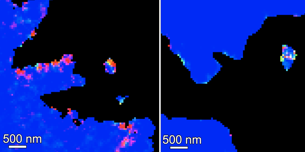
two bright colored images assign a color to the form of calcium present in coral skeletons. On the left there is a thicker band of non-blue (blue is crystalline aragonite) compared to the image on the right where there is almost all blue, indicating the skeleton on the right crystallizes to aragonite more quickly
Coral skeletons were studied with PEEM spectromicroscopy, which identifies the calcium spectrum associated with each imaging pixel, then renders it in false color depending on the form of calcium. Blue is aragonite, the insoluble, crystalline form of calcium carbonate; the other colors represent one of the two amorphous precursor forms, a mix of the two, or a mix of aragonite and precursor form. Acropora spp. (left), has more non-blue pixels compared to Turbinaria spp. (right), indicating that Acropora has more of the soluble, non-crystalline form in its growing skeleton. | Pupa Gilbert and team in JACS

The thicker the band of uncrystallized minerals, the slower the crystallization process.

“If the surface of the coral skeleton, where all this amorphous calcium carbonate is being deposited by the living animal, crystallizes quickly, then that particular species is resilient to ocean acidification; if it crystallizes slowly, then it’s vulnerable,” Gilbert says. “For once, it’s a really simple mechanism.”

The mechanism may have worked out to be simple, but the data analysis required to process and interpret the PEEM images is anything but. Each pixel of imaging data acquired has a calcium spectrum that needs to be analyzed, which results in millions of data points. Processing the data includes many decision-making points, plus massive computing power.

The team has tried to automate the analysis or use machine-learning techniques, but those methods have not worked out. Instead, Gilbert has found that humans making decisions are the best data processors.

Gilbert did not want to base conclusions off the decision-making of just one or two people. So she hired a group of UW–Madison undergraduates, most of whom came from the Mercile J. Lee Scholars Program, which works to attract and retain talented students from underrepresented groups. This team provided a large and diverse group of decision makers.

a zoom screen showing several of the people who conducted the study
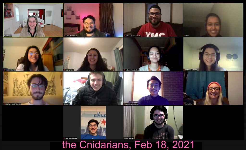
Gilbert and her research team met several times a week via Zoom, where students were assigned the same dataset to process in parallel and discuss at their next meeting. The Cnidarians — named after the phylum to which corals belong — include current and former UW–Madison undergraduates: Celeo Matute Diaz, Jorge Rivera Colon, Asiya Ahmed, Virginia Quach, Gabi Barreiro Pujol, Isabelle LeCloux, Sydney Davison, Connor Klaus, Jaden Sengkhamee, Evan Walch and Benjamin Fordyce; and graduate students Cayla Stifler, and Connor Schmidt. Schmidt was also the lead author of the study. | Provided by Pupa Gilbert

Dubbed the Cnidarians — from the phylum to which corals, anemones and jellyfish belong — this group of students became integral members of the team. They met several times a week via Zoom, when Gilbert would assign multiple students the same dataset to process in parallel and discuss at their next meeting.

“If multiple people come up with precisely the same solution even though they make different decisions, that means our analysis is robust and reliable,” Gilbert says. “The Cnidarians’ contributions were so useful that 13 of them are co-authors on this study.”

THIS STUDY WAS SUPPORTED BY THE DEPARTMENT OF ENERGY (DE-FG02-07ER15899 AND DE-AC02-05CHH11231), THE NATIONAL SCIENCE FOUNDATION (DMR-1603192) AND THE EUROPEAN RESEARCH COUNCIL (755876).

Bucket brigades and proton gates: Researchers shed new light on water’s role in photosynthesis

This story is adapted from one originally published by SLAC by Ali Sundermier

Photosystem II is a protein in plants, algae and cyanobacteria that uses sunlight to break water down into its atomic components, unlocking hydrogen and oxygen. A longstanding question about this process is how water molecules are funneled into the center of Photosystem II, where water is split to produce the oxygen we breathe. A better understanding of this process could inform the next generation of artificial photosynthetic systems that produce clean and renewable energy from sunlight and water.

In a paper published last week in Nature Communications, an international collaboration between scientists at the Department of Energy’s Lawrence Berkeley National Laboratory (LBNL), SLAC National Accelerator Laboratory and several other institutions uncovers how the protein takes in water and how hydrogen is removed in order to release the oxygen molecules.

Profile picture of Uwe Bergmann
Uwe Bergmann

“Plants use the energy from sunlight to split two water molecules and produce the oxygen we breath. The study shows for the first time atomic-resolution snapshots of the likely channel and gate, where the water molecules arrive to the catalytic center to be split apart, and the channel where the protons are shuttled out during the splitting,” says Uwe Bergmann, the Martin L. Perl professor in ultrafast x-ray science at UW–Madison. “This information will help our understanding of one of the most fundamental reactions on earth, and how we might use sunlight in the future to create fuels.”

At SLAC’s Linac Coherent Light Source (LCLS) X-ray laser, the team illuminated samples from cyanobacteria with ultrafast pulses of X-rays to collect both X-ray crystallography and spectroscopy data to simultaneously map the protein structure and how electrons flow in the protein. Through this technique, they are able to test competing theories of how Photosystem II splits water into oxygen. Over the past few years, the team has used this method to observe various steps of this water-splitting cycle at the temperature at which it occurs in nature. 

Scientists at UW–Madison have been instrumental to developing these and related x-ray imaging methods over the last decade.

The center of the protein acts as a catalyst, which drives certain chemical reactions to happen in a highly efficient manner. This research seeks to unlock how nature has optimized this catalytic process over millions of years of evolution. A cluster of four manganese atoms and one calcium atom are connected by oxygen atoms, and surrounded by water and the outer layers of the protein. In the step the scientists looked at, water flows through a pathway into the center of the protein, where one water molecule ultimately forms a bridge between a manganese atom and a calcium atom. The researchers showed that this water molecule likely provides one of the oxygen atoms in the oxygen molecule produced at the end of the cycle.

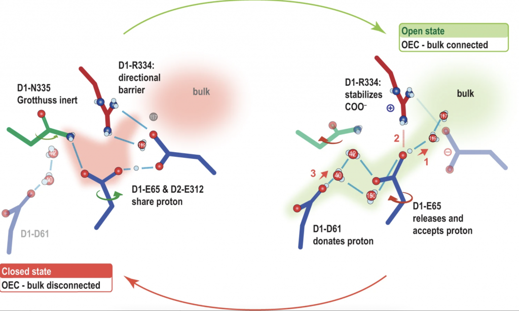
a schematic of the proposed mechanism is shown
The proposed proton gate around D1-E65, D2-E312, and D1-R334 in the open and closed state. | In Nature Communications, https://doi.org/10.1038/s41467-021-26781-z

Last year, the researchers discovered that Photosystem II ferries water into the center as if through a bucket brigade: Water molecules move in many small steps from one end of the pathway to the other. They also showed that the calcium atom within the center could be involved in shuttling the water in. In this most recent study, the researchers pinpoint, for the first time, the exact pathway where this process unfolds.

“This might prevent water from interacting with the center prematurely, resulting in unwanted intermediates such as peroxide that can cause damage to the enzyme,” said Jan Kern, staff scientist at LBNL and one of the corresponding authors.

The researchers also showed that there is another pathway dedicated to removing hydrogen protons generated during the water-splitting reaction. In the proton pathway, they discovered the existence of a “proton gate,” which blocks the proton from coming back to the center.

“These results show where and how the water molecules enter the catalytic site, and where the protons are released, advancing our understanding of how two waters may come together to form the oxygen we breathe,” said Junko Yano, senior scientist at LBNL and one of the corresponding authors. “It demonstrates that it is just not enough to determine the structure of the main catalytic center, but it is also important to understand how the entire protein carries out the reaction.”

In addition to SLAC and LBNL, the collaboration includes researchers from Uppsala University in Sweden; Humboldt University of Berlin; and the University of Wisconsin-Madison.

LCLS is a DOE Office of Science user facility. This research was supported by the Office of Science.


New nondestructive optical technique reveals the structure of mother-of-pearl

Most people know mother-of-pearl, an iridescent biomineral also called nacre, from buttons, jewelry, instrument inlays and other decorative flourishes. Scientists, too, have admired and marveled at nacre for decades, not only for its beauty and optical properties but because of its exceptional toughness.

“It’s one of the most-studied natural biominerals,” says Pupa Gilbert, a University of Wisconsin–Madison physics professor who has studied nacre for more than a decade. “It may not look like much — just a shiny, decorative material. But it can be 3,000 times more resistant to fracture than aragonite, the mineral from which it’s made. It has piqued the interest of materials scientists because making materials better than the sum of their parts is extremely attractive.”

Now, a new, nondestructive optical technique will unlock even more knowledge about nacre, and in the process could lead to a new understanding of climate history. Gilbert, UW–Madison electrical engineering professor Mikhail Kats — who is also an affiliate professor of physics — their students, and collaborators described the technique, called hyperspectral interference tomography, today in the journal Proceedings of the National Academy of Sciences.

Read the Full News Story | PNAS study

Pupa Gilbert elected Fellow of the Mineralogical Society of America

Proflie photo of Pupa Gilbert

Congrats to Prof. Pupa Gilbert on her election as a Fellow of the Mineralogical Society of America! Members who have contributed significantly to the advancement of mineralogy, crystallography, geochemistry, petrology, or allied sciences and whose scientific contribution utilized mineralogical studies or data, may be designated as Fellows upon proper accreditation by the Committee on Nomination for Fellows and election by the Council. The number of fellows elected each year cannot exceed 0.5% of MSA membership.

Fellows newly elected in 2020 are Jeffrey Catalano, Sylvie Demouchy, Pupa Gilbert, Jun-ichi Kimura, Othmar Muntener, Marc Norman, Alison Pawley, Mark Rivers, Ian Swainson, and Takashi Yoshino.

Full list of MSA Fellows

A better understanding of coral skeleton growth suggests ways to restore reefs

Coral reefs are vibrant communities that host a quarter of all species in the ocean and are indirectly crucial to the survival of the rest. But they are slowly dying — some estimates say 30 to 50 percent of reefs have been lost — due to climate change.

In a new study, University of Wisconsin–Madison physicists observed reef-forming corals at the nanoscale and identified how they create their skeletons. The results provide an explanation for how corals are resistant to acidifying oceans caused by rising carbon dioxide levels and suggest that controlling water temperature, not acidity, is crucial to mitigating loss and restoring reefs.

“Coral reefs are currently threatened by climate change. It’s not in the future, it’s in the present,” says Pupa Gilbert, a physics professor at UW–Madison and senior author of the study. “How corals deposit their skeletons is fundamentally important to assess and help their survival.”

Read the Full Story | Link to the PNAS study