Coral skeleton formation rate determines resilience to acidifying oceans

A new University of Wisconsin–Madison study has implications for predicting coral reef survival and developing mitigation strategies against having their bony skeletons weakened by ocean acidification.

Though coral reefs make up less than one percent of the ocean floor, these ecosystems are among the most biodiverse on the planet — with over a million species estimated to be associated with reefs.

The coral species that make up these reefs are known to be differently sensitive or resilient to ocean acidification — the result of increasing atmospheric carbon dioxide levels. But scientists are not sure why.

In the study, researchers show that the crystallization rate of coral skeletons differs across species and is correlated with their resilience to acidification.
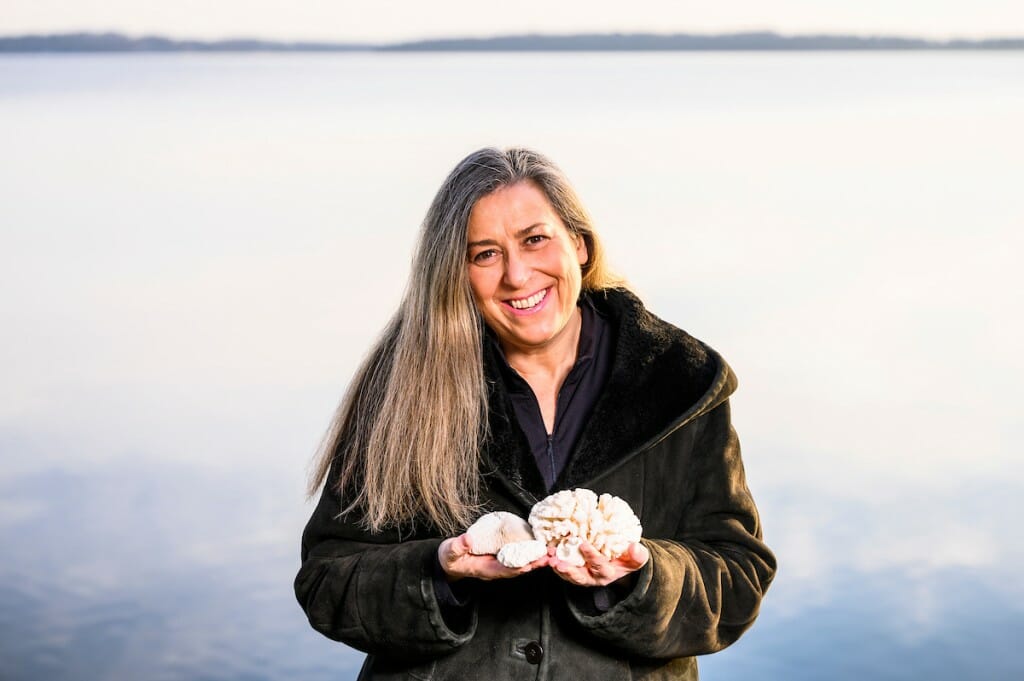
A woman holding two coral species stands in front of a body of water
“Finding solutions that are science-based is a priority,” says physics professor Pupa Gilbert, shown here with samples of scleractinian coral along the Lake Monona shoreline in Madison. | Photo: Jeff Miller

“Many agencies keep putting out reports in which they say, ‘Yes, coral reefs are threatened,’ with no idea what to do,” says Pupa Gilbert, a physics professor at UW–Madison and senior author of the study that was published Jan. 17 in the Journal of the American Chemical Society. “Finding solutions that are science-based is a priority, and having a quantitative idea of exactly what’s happening with climate change to coral reefs and skeletons is really important.”

Reef-forming corals are marine animals that produce a hard skeleton made up of the mostly insoluble crystalline material aragonite. Aragonite forms when precursors made up of a more soluble form, amorphous calcium carbonate, are deposited onto the growing skeleton and then crystallize.

The team studied three genera of coral and took an in-depth look at the components of their growing skeletons. They used a technique that Gilbert pioneered called PEEM spectromicroscopy, which detects the different forms of calcium carbonate with the greatest sensitivity to date.

When they used these spectromicroscopy images to compare the thickness of amorphous precursors to the crystalline form, they found that Acropora, which is more sensitive to acidification, had a much thicker band of amorphous calcium carbonate than Stylophora, which is less sensitive.

A third genus of unknown sensitivity, Turbinaria, had an even thinner amorphous precursor layer than Stylophora, suggesting it should be the most resilient of the three to ocean acidification.
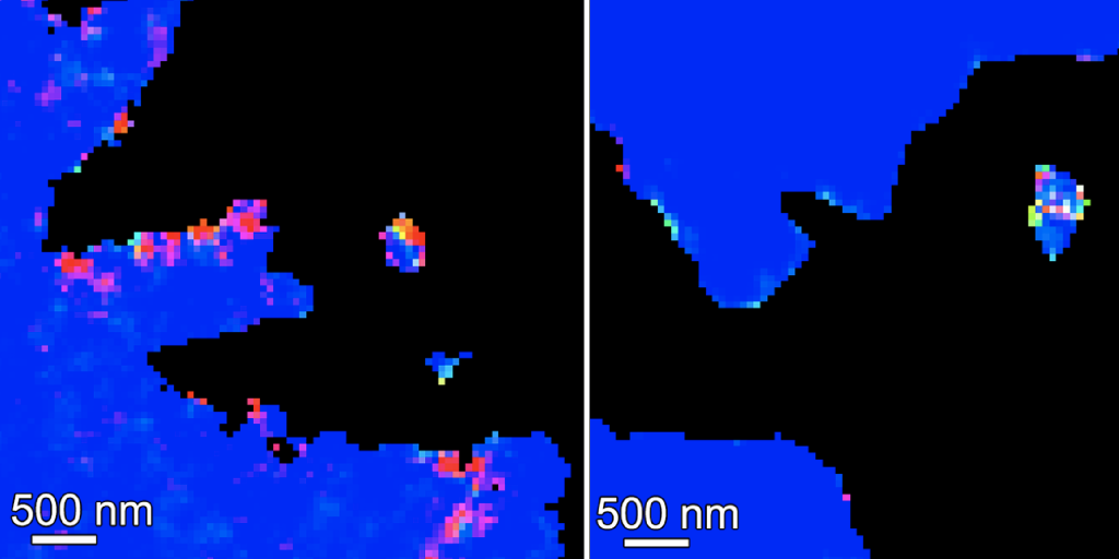
two bright colored images assign a color to the form of calcium present in coral skeletons. On the left there is a thicker band of non-blue (blue is crystalline aragonite) compared to the image on the right where there is almost all blue, indicating the skeleton on the right crystallizes to aragonite more quickly
Coral skeletons were studied with PEEM spectromicroscopy, which identifies the calcium spectrum associated with each imaging pixel, then renders it in false color depending on the form of calcium. Blue is aragonite, the insoluble, crystalline form of calcium carbonate; the other colors represent one of the two amorphous precursor forms, a mix of the two, or a mix of aragonite and precursor form. Acropora spp. (left), has more non-blue pixels compared to Turbinaria spp. (right), indicating that Acropora has more of the soluble, non-crystalline form in its growing skeleton. | Pupa Gilbert and team in JACS

The thicker the band of uncrystallized minerals, the slower the crystallization process.

“If the surface of the coral skeleton, where all this amorphous calcium carbonate is being deposited by the living animal, crystallizes quickly, then that particular species is resilient to ocean acidification; if it crystallizes slowly, then it’s vulnerable,” Gilbert says. “For once, it’s a really simple mechanism.”

The mechanism may have worked out to be simple, but the data analysis required to process and interpret the PEEM images is anything but. Each pixel of imaging data acquired has a calcium spectrum that needs to be analyzed, which results in millions of data points. Processing the data includes many decision-making points, plus massive computing power.

The team has tried to automate the analysis or use machine-learning techniques, but those methods have not worked out. Instead, Gilbert has found that humans making decisions are the best data processors.

Gilbert did not want to base conclusions off the decision-making of just one or two people. So she hired a group of UW–Madison undergraduates, most of whom came from the Mercile J. Lee Scholars Program, which works to attract and retain talented students from underrepresented groups. This team provided a large and diverse group of decision makers.
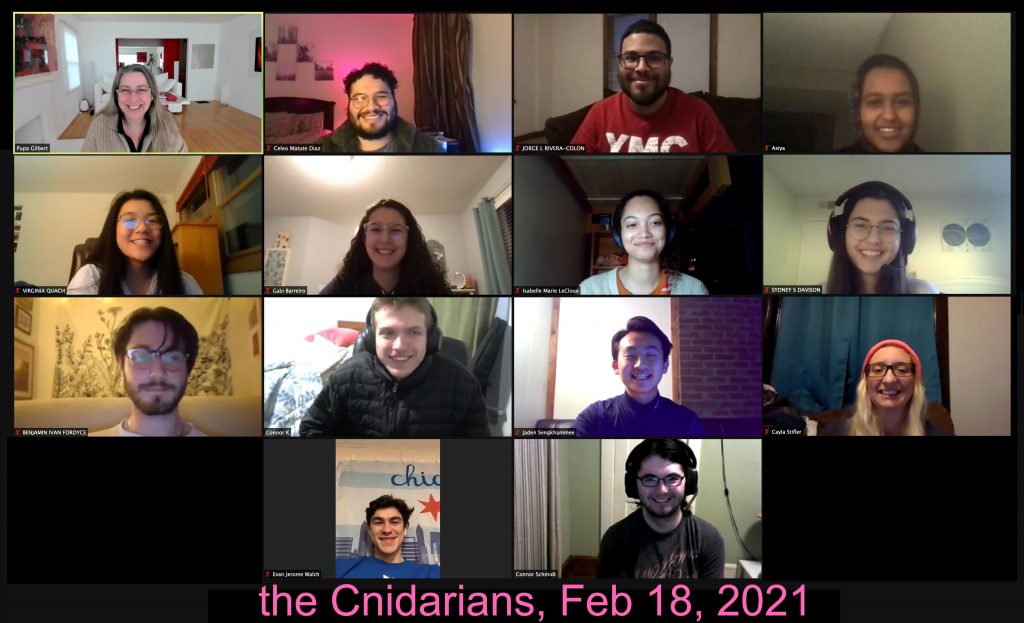
a zoom screen showing several of the people who conducted the study
Gilbert and her research team met several times a week via Zoom, where students were assigned the same dataset to process in parallel and discuss at their next meeting. The Cnidarians — named after the phylum to which corals belong — include current and former UW–Madison undergraduates: Celeo Matute Diaz, Jorge Rivera Colon, Asiya Ahmed, Virginia Quach, Gabi Barreiro Pujol, Isabelle LeCloux, Sydney Davison, Connor Klaus, Jaden Sengkhamee, Evan Walch and Benjamin Fordyce; and graduate students Cayla Stifler, and Connor Schmidt. Schmidt was also the lead author of the study. | Provided by Pupa Gilbert

Dubbed the Cnidarians — from the phylum to which corals, anemones and jellyfish belong — this group of students became integral members of the team. They met several times a week via Zoom, when Gilbert would assign multiple students the same dataset to process in parallel and discuss at their next meeting.

“If multiple people come up with precisely the same solution even though they make different decisions, that means our analysis is robust and reliable,” Gilbert says. “The Cnidarians’ contributions were so useful that 13 of them are co-authors on this study.”

THIS STUDY WAS SUPPORTED BY THE DEPARTMENT OF ENERGY (DE-FG02-07ER15899 AND DE-AC02-05CHH11231), THE NATIONAL SCIENCE FOUNDATION (DMR-1603192) AND THE EUROPEAN RESEARCH COUNCIL (755876).

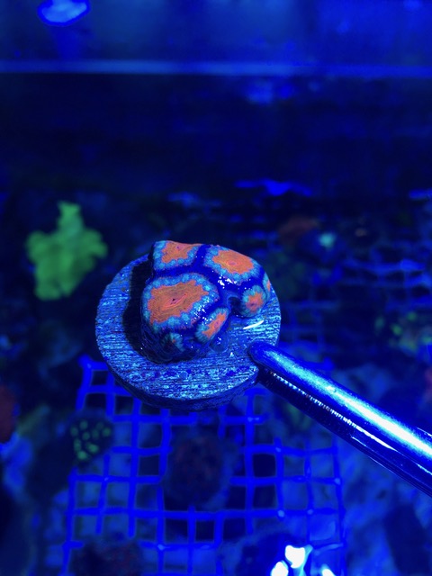
New nondestructive optical technique reveals the structure of mother-of-pearl

Most people know mother-of-pearl, an iridescent biomineral also called nacre, from buttons, jewelry, instrument inlays and other decorative flourishes. Scientists, too, have admired and marveled at nacre for decades, not only for its beauty and optical properties but because of its exceptional toughness.

“It’s one of the most-studied natural biominerals,” says Pupa Gilbert, a University of Wisconsin–Madison physics professor who has studied nacre for more than a decade. “It may not look like much — just a shiny, decorative material. But it can be 3,000 times more resistant to fracture than aragonite, the mineral from which it’s made. It has piqued the interest of materials scientists because making materials better than the sum of their parts is extremely attractive.”

Now, a new, nondestructive optical technique will unlock even more knowledge about nacre, and in the process could lead to a new understanding of climate history. Gilbert, UW–Madison electrical engineering professor Mikhail Kats — who is also an affiliate professor of physics — their students, and collaborators described the technique, called hyperspectral interference tomography, today in the journal Proceedings of the National Academy of Sciences.

Read the Full News Story | PNAS study

Pupa Gilbert elected Fellow of the Mineralogical Society of America

Proflie photo of Pupa Gilbert

Congrats to Prof. Pupa Gilbert on her election as a Fellow of the Mineralogical Society of America! Members who have contributed significantly to the advancement of mineralogy, crystallography, geochemistry, petrology, or allied sciences and whose scientific contribution utilized mineralogical studies or data, may be designated as Fellows upon proper accreditation by the Committee on Nomination for Fellows and election by the Council. The number of fellows elected each year cannot exceed 0.5% of MSA membership.

Fellows newly elected in 2020 are Jeffrey Catalano, Sylvie Demouchy, Pupa Gilbert, Jun-ichi Kimura, Othmar Muntener, Marc Norman, Alison Pawley, Mark Rivers, Ian Swainson, and Takashi Yoshino.

Full list of MSA Fellows

A better understanding of coral skeleton growth suggests ways to restore reefs

Coral reefs are vibrant communities that host a quarter of all species in the ocean and are indirectly crucial to the survival of the rest. But they are slowly dying — some estimates say 30 to 50 percent of reefs have been lost — due to climate change.

In a new study, University of Wisconsin–Madison physicists observed reef-forming corals at the nanoscale and identified how they create their skeletons. The results provide an explanation for how corals are resistant to acidifying oceans caused by rising carbon dioxide levels and suggest that controlling water temperature, not acidity, is crucial to mitigating loss and restoring reefs.

“Coral reefs are currently threatened by climate change. It’s not in the future, it’s in the present,” says Pupa Gilbert, a physics professor at UW–Madison and senior author of the study. “How corals deposit their skeletons is fundamentally important to assess and help their survival.”

Read the Full Story | Link to the PNAS study